Why do we need microscopes?
- Cells are tiny, so light microscopes are used to see their details.
- A slide is a thin piece of glass used to hold objects which are examined under a microscope. are used to examine objects under the microscope.
- Stains are used to help see different structures in the cell.
- Biological drawings can be done from observations made with microscopes.
How to make a temporary slide
Plant cells:
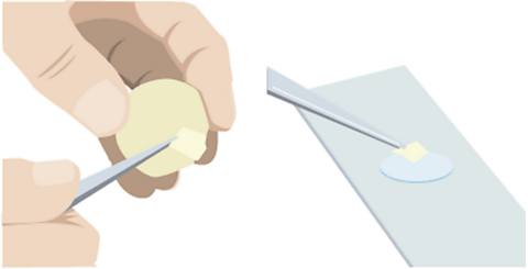
Peel a thin layer of cells from an onion.
Place on a slide.
Add a drop of iodine (a chemical stain).
Gently lower a coverslip to avoid air bubbles and prevent drying.
Animal cells:
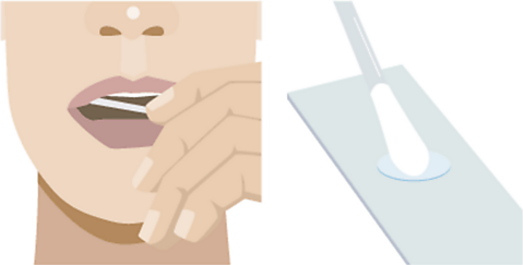
Collect cheek cells with a cotton bud.
Smear onto a slide.
Add methylene blue (a chemical stain).
Gently lower a coverslip to avoid air bubbles.
Chemical stains highlight cell parts making them easier to see. It’s temporary as the cells are not preserved so will rot.
Observing and recording
Using a light microscope
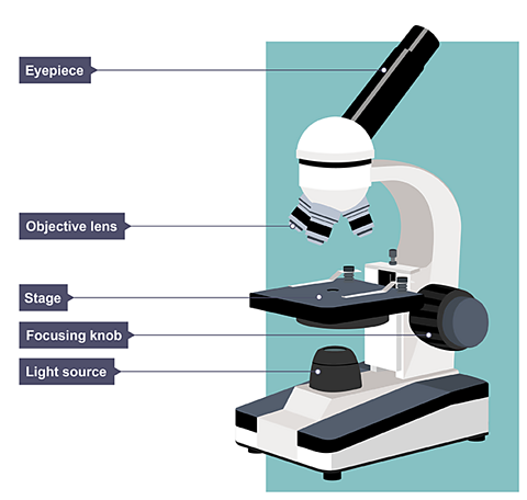
When using a light microscope, start with the low power objective lens as the field of view will be wider, increasing the number of cells you are able to see, making it easier to locate cells.
Once centred, switch to a higher power lens for detailed viewing.
The lens is very close to the slide so be careful to avoid damaging it.
Total magnification = eyepiece magnification × objective lens magnification.
| Objective lens | Eyepiece magnification | Total magnification | |
|---|---|---|---|
| Low power | 4X | 10X | 40X |
| Medium power | 10X | 10X | 100X |
| High power | 40X | 10X | 400X |
Drawing cell structures:
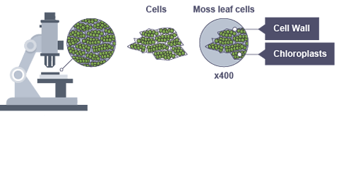
A good biological drawing should be:
drawn in pencil with firm, continuous lines (no sketching)
large and proportionate to the observed cell
clearly labelled
given a title with magnification or size
Quiz time!
Watch: The power of the microscope
You might not think that a tube with a couple of bits of glass in it could have completely changed the world, but the microscope did just that.
When it was invented, in the 1600s, it kicked off the start of modern biology, of understanding the structures of life.
Today, microscopes are much more powerful than those first devices, but optical microscopes still work on essentially the same principles. They pass light through glass lenses, in order to magnify the specimen.
A really powerful professional model can get up to around 2,000 times magnification which means it's making an object look 2,000 times larger than it actually is. Now, that's powerful enough to be able to see an animal cell in detail.
Images look like this because we often stain the cells so that we can see the organelles more clearly. Or we can see the plant cells which make up a leaf, and it's just about powerful enough to see bacteria.
Ok, so the more magnification we can get from a microscope the better it is. But it's not just size that matters, it’s also resolution. Resolution is the amount of detail that can be seen in the image and it's measured by the smallest distance between 2 points that can clearly be distinguished from one another.
And, if you're wondering what the boss of the microscope world is, something that packs a punch with both magnification and resolving power, look no further than the electron microscope.
Electron microscopes don't use light at all, so they're not optical microscopes. And they don't use glass lenses either. Instead they pass a beam of electrons through a sample, and then by detecting how many electrons pass through the sample, they build up an image. An incredibly detailed image.
With electron microscopes it's possible to not only see an animal cell, but to actually zoom in and see the detailed internal structures of organelles like the nucleus in an animal cell or the chloroplasts in plant cells. You can even see the plasmids inside a bacterium!
Electron microscopes let us see things hundreds of thousands, or even millions of times bigger than they really are! But I’m not done yet. Special scanning electron microscopes can take us further than a flat 2D image. This is a 3D image of a flea!
I should mention, there are disadvantages to electron microscopes as well though. First, for many models, the specimens have to have a thin coating of metal added before they are put in a vacuum chamber. Second, they're big machines, so you can't easily move them around, and, third, they cost serious money. That's why the good old optical microscope is still used for a lot of research work, and in schools.
Those first inventors can't have known what they'd started and the incredible developments their optical microscopes would lead to.
The microscope has enabled us to understand how living things work more than almost any other scientific tool.